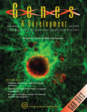
A new paper by Crislyn D’Souza-Schorey, professor of biological sciences at the University of Notre Dame, discusses the biology of tumor-derived microvesicles and their clinical application as circulating biomarkers. Microvesicles are membrane-bound sacs released by tumor cells and can be detected in the body fluids of cancer patients.
The World Health Organization estimates that cancer will cause approximately 9 million deaths in 2015. The rising prevalence of the disease is a major factor that drives the growth of the oncology biomarkers market. Biomarkers can be defined as any biological, chemical or physical parameter that can be utilized as an indicator of physiological or disease status. Thus, biomarkers are useful in cancer screening and detection and drug design and also in boosting the effectiveness of cancer care by allowing physicians to tailor therapies for individual patients — an approach known as personalized medicine.
The new paper discusses the potential of microvesicles to present a combination of disease- and tissue-specific markers that would constitute a unique and identifiable biosignature for individual cancers.
“As such, it would make their sampling over time a preferred method to monitor changes to the tumor in response to treatment, especially for tissues such as the ovary or pancreas, where repeated biopsies of these organs is impractical,” D’Souza-Schorey said.
Profiling of microvesicles could form the basis of personalized, targeted cancer therapies, especially as more reliable and rapid profiling technologies become available.

“For example, certain markers like HER2/neu, in addition to being elevated in breast cancer, is also increased in a relatively smaller subset of other cancers such as ovarian cancer,” D’Souza-Schorey said. “This latter group of patients would benefit from existing treatment strategies that target the HER2 receptor.”
The approach could be advantageous over currently used approaches of profiling whole tissue or un-fractionated body fluid particularly if circulating microvesicles indeed concentrate molecular changes that occur in the tumor, as it would increase the sensitivity of detecting critical markers of cancer progression.
“One complicating factor, though, is the presence of shed vesicles from other non-tumor cell types also in direct contact with these body fluids,” D’Souza-Schorey said. “Thus, equally significant is the development of strategies to selectively capture tumor-specific markers that separate from other shed vesicle populations.”
In collaboration with local oncologists, the D’Souza-Schorey laboratory is investigating the potential of microvesicles as a cancer diagnostic platform, a project under the umbrella of Notre Dame’s Advanced Diagnostics and Therapeutics Initiative. The lab’s research on the biology of microvesicles and their roles in tumor progression is supported by the National Cancer Institute and the Indiana Clinical and Translational Sciences Institute.
“Despite considerable strides, effort and investment in cancer biomarker research in the past decade, there are still more desirable outcomes, most especially enhanced sensitivity to enable early detection,” D’Souza-Schorey said. “An effective biomarker platform that will overcome these challenges would be paradigm-shifting in cancer care.”
The paper, which appears in the June 15 issue of the journal Genes and Development, was coauthored by Notre Dame graduate student James Clancy.
Contact: Crislyn D’Souza-Schorey, 574-631-3735, cdsouzas@nd.edu