|
|
|
Free Neuropathology 1:7 (2020) |
|
Review |
|
Dementia with Lewy bodies – a clinicopathological update |
|
János Bencze1,2, Woosung Seo3, Abdul Hye4,5, Dag Aarsland5,6,7, Tibor Hortobágyi2,6,7,8 |
|
1 Department of Pathology, Faculty of Medicine, University of Debrecen, Debrecen, Hungary |
|
Corresponding author: |
|
Submitted: 31 December 2019 Accepted: 06 February 2020 Published: 18 February 2020 |
|
Keywords: α-synuclein, Biomarkers, Diagnostic criteria, Clinico-pathological correlation, Dementia with Lewy bodies |
|
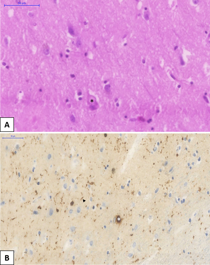
Abstract Dementia is one of the major burdens of our aging society. According to certain predictions, the number of patients will double every 20 years. Although Alzheimer’s disease (AD), as the most frequent neurodegenerative dementia, has been extensively analysed, less is known about dementia with Lewy bodies (DLB). Neuropathological hallmarks of DLB are the deposition of intracellular Lewy bodies (LB) and Lewy neurites (LN). DLB belongs to the α-synucleinopathies, as the major component of these inclusions is pathologically aggregated α-synuclein. Depending on the localisation of LBs and LNs in the central nervous system cognitive and motor symptoms can occur. In our work, we will systematically review the possible etiology and epidemiology, pathological (both macroscopic and microscopic) features, structural and functional imaging findings, with a special emphasis on the clinico-pathological correlations. Finally, we summarize the latest clinical symptoms-based diagnostic criteria and the novel therapeutic approaches. Since DLB is frequently accompanied with AD pathology, highlighting possible differential diagnostic approaches is an integral part of our paper. Although our present knowledge is insufficient, the rapid development of diagnostic and research methods provide hope for better diagnosis and more efficient treatment, contributing to a better quality of life. Abbreviations AA, Alzheimer’s Association; AD, Alzheimer’s disease; Aβ, Amyloid-beta; APOE, Apolipoprotein E gene; BNE, BrainNet Europe Consortium; BOLD, Blood oxygen level dependent; ChAT, Choline-acetyltransferase; ChAT-I, Acetyl-cholinesterase inhibitors; CR, Creatinine; CSF, Cerebrospinal fluid; DLB, Dementia with Lewy bodies; DMN, Default mode network; DSM, Diagnostic Statistical Manual; fMRI, Functional MRI; FP-CIT, [123I] 2ß-carbomethoxy-3b-(4-iodophenyl)-N-(3-fluoropropyl) nortropane; GBA, Glucosylceramidase-beta gene; iLBD, Incidental Lewy body disease; LB, Lewy body; LBDA, Lewy Body Dementia Association; LN, Lewy neurite; MAPT, Microtubule associated protein tau gene; MDS, Movement Disorders Society; MRI, Magnetic resonance imaging; NAA, N-acetyl aspartate; NCD, Neurocognitive disorder; NF, Neurofilament; NIA, National Institute on Aging; NMDAR, N-methyl-D-aspartate receptor; PD, Parkinson’s disease; PDD, Parkinson’s disease dementia; PET, Positron emission tomography; PSD95, Density protein 95; REM, Rapid-eye-movement; SCARB2, Scavenger Receptor Class B Member 2; SNARE, SNAP (Soluble NSF Attachment Protein) Receptor; SNCA, α-synuclein gene; SPECT, Single-photon emission computed tomography; SSRI, Selective serotonin reuptake inhibitors; UPR, Unfolded protein response; WML, White matter lesions; ZnT3, Zinc transporter 3 Epidemiology and etiology Dementia with Lewy bodies (DLB) is the second most common primary neurodegenerative dementia. The etiology is mainly unknown, however, in certain cases there is strong evidence of genetic background. A few papers have reported families with accumulating occurrence of cognitive impairment throughout generations1. The vast majority of the familial cases are traced to α-synuclein (SNCA) gene alterations, particularly to E46K mutation2. Five candidate genes, including APOE, GBA, MAPT, SNCA and SCARB2, are considered as a significant risk factor for DLB, nevertheless, further genetic studies are needed3. Although it mostly appears in older age, dementia is not strictly associated with aging. Rarely it may occur under the age of 65 and even in young adulthood. Surprisingly in the case report of a teenager, the clinical symptoms and post-mortem pathological findings were consistent with those of in DLB4. The prevalence of the disease is not clearly established. According to a comprehensive analysis of epidemiological data, the prevalence is 4.2% in community based and 7.5% in clinical studies, while the incidence is 3.8% and grows linearly with aging5 and the prevalence of DLB among patients with dementia is probably around 15%6. Pathologic background 1. Macroscopic observation Many of the general pathologic features resemble those in Parkinson’s disease (PD). The brain weight is often within the normal limits, mild cortical atrophy of the frontal lobe, neuromelanin pigment loss in the substantia nigra and locus coeruleus are noted. In the case of severe concomitant AD pathology, atrophy of the temporal and parietal regions is more evident. In contrast to AD, the general brain atrophy is less prominent in DLB along with the relatively preserved temporal lobe and hippocampus7. 2. Microscopic feature Lewy bodies (LBs), the key pathological findings in DLB, were initially described by Friedrich Lewy in 19128. However, LBs are characteristic of other neurodegenerative diseases including PD. Their typical appearance, stained with conventional hematoxylin-eosin, is a central spherical eosinophilic core surrounded by a peripheral halo situated intracellularly, causing dislocation of subcellular organs. These features are regularly found in brainstem predominant LB formation. The rest of the brain expresses rather irregular LBs without the typical peripheral halo (Figure 1).
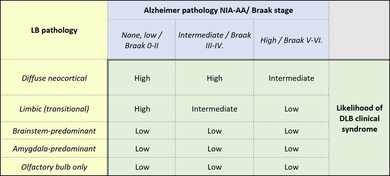
Figure 1: Dementia with Lewy bodies (DLB) specific pathological changes are shown with hematoxylin and eosin (HE) staining (Panel A) and α-synuclein immunohistochemistry (ICH) (Panel B). Cortical-type Lewy bodies (LBs) are eosinophilic intracellar neuronal inclusions which dislocate the nucleus (Panel A, black star). α-synuclein IHC highlights LBs (white star, Panel B) and LNs (arrowhead, Panel B). Histopathologically, the dominant component of the central core is α-synuclein, while the peripheral halo consists of several ubiquitinated proteins9. Lewy neurites (LNs), as an additional hallmark of α-synucleinopathies, are abnormal thickened neurites containing filaments corresponding to those in LBs10. Interestingly, in experimental mouse models mutant SNCA inoculation of wild type mouse causes LB/LN-like pathology, supporting the pathognomonic role of the protein11. The background of LB genesis is still undiscovered, although numerous hypotheses have been proposed trying to explain the proper mechanism. According to the aggresome hypothesis, LB formation is originally a neuroprotective process, facilitating the cells to remove harmful proteins12. However, failure of the aggresome formation may occur, leading to excessive LB development13. Other authors presume that autophagy dysfunction is responsible for the neuronal loss14. A recent study suggests that the increased unfolded protein response (UPR) activation has an important role in the LB pathology15. Nevertheless, according to Tompkins et al. neurons burdened with LBs are less apoptotic16. Moreover, the downregulation of tyrosine hydroxylase enzymes protects against toxic products of dopamine oxidation17. These findings confirm the theory that α-synuclein is physiologically involved in the maintenance of cell homeostasis. The first standardized criterion assessing the connections between pathological findings and DLB was made by Kosaka et al. in 1984. Based on the anatomical distribution of pathology they determined three different subtypes: i) brainstem predominant LBs (commonly in PD) ii) limbic (transitional) LBs and iii) diffuse cortical LBs18. Later the Consortium on DLB International Workshop has improved the original assignment and published a more detailed instruction emphasizing the importance of the diagnostic procedure. The widely used immunohistochemical markers, α-synuclein (the most specific) and p62 serve the accurate pathological diagnosis19. The latest McKeith diagnostic consensus criteria recommends a semiquantitative grading of 10 different brain regions (dorsal motor nucleus of Vagus, locus coeruleus, substantia nigra, nucleus basalis of Meynert, amygdala, transentorhinal and cingulate gyri, temporal-, frontal- and parietal lobes) based on the lesion density instead of the previously used LB counting method. Moreover, it suggests two additional categories, the amygdala-predominant and the olfactory bulb only DLB. According to the distribution of LB pathology on α-synuclein immunostained slides, the scoring system distinguishes four stages: 1 – mild (sparse LBs or LNs); 2 – moderate (1< LBs in a low power field and sparse LNs); 3 - severe (4≤ LBs and scattered LNs in a low power field); 4 – very severe (numerous LBs and numerous LNs). Finally, as a synthesis of score and localization it ranks the seen pathology into one out of the three previously mentioned subtypes20. Besides the McKeith staging and subtyping, the other widely used evaluating technique is the Braak staging21. The authors proposed to assess the severity of Lewy-type pathology labelled by α-synuclein immunostaining in 13 different brain areas (dorsal motor nucleus of Vagus, locus coeruleus, raphe, substantia nigra, CA2 region of hippocampus, nucleus basalis of Meynert, transentorhinal-, cingulate- and insular gyri, temporo-occipital-, temporal-, frontal- and parietal lobes). Considering that neither McKeith nor Braak protocol could reach more than 80% inter-observer agreement; the original methods have been modified by Leverenz et al.22 and Müller et al.23, respectively. However, their results did not lead to a significant improvement in the scoring systems. Thus, in 2009 the BrainNet Europe Consortium (BNE) revised their former assignments and suggested modifications, to reach a better inter-observer agreement. Using the original McKeith and Braak staging, but eliminating their major pitfalls and obstacles as well as introducing the amygdala predominant category, BNE’s novel strategy resulted in above 80% agreement in both typing and staging of α-synuclein pathology24. Beach et al. also published their unified staging system with the aim of dividing every subject with Lewy-type α-synuclein pathology into a well-defined neuropathological group25. In 2009 they used McKeith26 and Braak21 staging systems and failed to categorize individuals with olfactory bulb or limbic-predominant LB pathology. Investigating 10 standard brain regions they could classify all patients with PD, DLB, incidental Lewy body disease (iLBD) and AD with concomitant LB pathology into one of the following stages: I Olfactory Bulb Only; IIa Brainstem-predominant; IIb Limbic-predominant; III Brainstem and Limbic; IV Neocortical. Moreover, they found strong correlation between the progression through these stages and the severity of nigrostriatal degeneration, cognitive impairment and motor dysfunction. It should be noted that olfactory bulb only and amygdala-predominant subtypes are also included in the current McKeith criteria20. As mentioned above, AD pathology is frequently encountered in DLB. Approximately 80% of patients have diffuse amyloid-beta (Aβ) plaques and 60% have neurofibrillary tangles with varying severity in the entorhinal cortex and rarely in the neocortex. Some of these cases meet the pathological criteria of AD27. In contrast, “pure” neuropathological form of DLB is less common. Autopsy series suggest that the frequency of this entity is approximately 25% of all DLB cases28,29. Theoretically, the likelihood to manifest DLB clinical syndrome is directly proportional to the severity of LB pathology and inversely proportional to the severity of AD pathology. The McKeith classification integrates the assessment of concomitant AD pathology by National Institute on Aging and Alzheimer’s Association (NIA-AA)30, the Braak criteria and the type of LB pathology20. Table 1 shows the probability of pathological findings in relation to DLB clinical syndrome. It is likely that not only the α-synuclein pathology is responsible for the cognitive decline, but plaques and phosphorylated tau proteins also contribute to the overall deficit31.
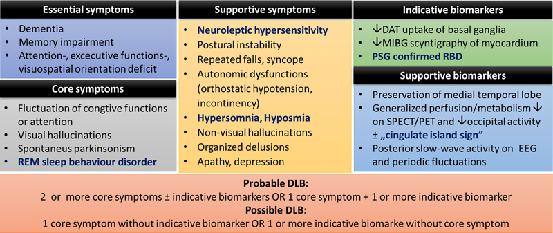
Table 1: Likelihood of Dementia with Lewy bodies (DLB) clinical syndrome resulting from the assessment of Alzheimer's-type and Lewy body pathology (LB= Lewy body; DLB=Dementia with Lewy bodies; NIA-AA = National Institute on Aging – Alzheimer’s Association) [Modified from McKeith et al.20] Cognitive impairment is also a common hallmark of DLB and Parkinson’s disease dementia (PDD). These two neurocognitive disorders share several clinical and neuropathological features, i.e. they are both characterized by cortical and subcortical α-synuclein/LB, β-amyloid and tau pathologies32. According to Jellinger et al., a common pathophysiology in DLB and PDD is synaptic dysfunction due to aggregation of α-synuclein in the presynapses. This results in disruption of axonal transport and neurotransmitter deprivation leading to neurodegeneration32. Interestingly, there are some morphologic differences as well. DLB seems to show higher load of β-amyloid and tau in several brain regions primarily in striatum, cortex, claustrum, amygdala and putamen compared to PDD32. Jellinger et al. also describes that α-synuclein distribution is different in DLB and PDD where α-synuclein load was highest in hippocampal subarea CA2 and in amygdala in DLB, whereas in PD it is highest in the cingulate cortex. Moreover, nigral neuronal loss is more marked in PDD which ultimately results in dopaminergic upregulation32. A simple interpretation of above findings could support the current diagnostic criteria where DLB is associated with early cognitive impairment including memory problems, which differs from PDD with mainly motor impairment in the early phase. In reality, it is not easy to define DLB based upon only the histological findings, thus in uncertain cases, anamnestic clinical data has invaluable support for the diagnostics. It is important to highlight that the listed strategies are not absolute or perfect diagnostic criteria, but rather useful schemes to predict the clinical syndrome of DLB based on the observed pathological findings. As mentioned above, a major weakness of current guidelines is the low inter-observer agreement; therefore further research is needed to assess the accuracy of the current clinical diagnostic criteria versus neuropathology similar to studies assessing the 2005 criteria in this respect33. 3. Synaptic alterations In connection with the dopaminergic neuronal loss of substantia nigra, dopamine transporter level is decreased in the striatum in DLB, although not to the same extent observed in PD34. The choline-acetyltransferase (ChAT) levels are lower than in patients with AD, who suffer from similarly severe dementia35,36. Interestingly, Dynamin-1, which takes part in the regulation of synaptic transmission, shows significantly decreased levels in the prefrontal cortex in parallel with the severity of cognitive decline37. In addition, the amount of zinc transporter 3 (ZnT3) and postsynaptic density protein 95 (PSD95), are significantly reduced in DLB compared to aged-controls and patients with AD38,39. It should be noted that at the beginning of the disease, an upregulation of soluble NSF attachment protein receptors (SNARE) complex is observed, probably as a compensatory response to the synaptic loss. However, during the progression of DLB synaptotagmin, synapsin and synaptophysin, proteins involved in the SNARE complex steadily disintegrate, contributing further to synaptic dysfunction40,41. Recent studies have revealed that more than 90% of α-synuclein aggregates are located at the presynapses leading to neurotransmitter deficits42,43. These findings suggest that degeneration of postsynaptic neurons may result from the loss of their inputs. This theory serves as a potential explanation for the DLB-specific clinical symptoms as well as raises the possibility of future curative treatments by pharmaceutical modification of neurotransmission. 4. Incidental Lewy-body disease (iLBD) In iLBD, patients have histologically detectable α-synuclein deposits in their brain, without presenting any clinical symptoms. Researchers accept that iLBD, which appears in approximately 8-17% of the clinically normal 60+ years old patients, is the early stage of PD or DLB44,45. Confirming this hypothesis, it is frequently observed in patients who represent the prodromes of PD, including olfactory dysfunction or bowel frequency46,47. The presence of neuronal loss is usually minimal in these cases contrary to PD or DLB48. Distribution of α-synuclein pathology in iLBD is particularly predictive, i.e. the brainstem predominant subtype frequently results in PD, while the cortical predominant subtype often leads to DLB44. Clinical symptoms The new Diagnostic Statistical Manual (DSM-5) (published in 2013) created a new neurocognitive disorder (NCD) group, replacing the former ‘dementia, delirium, amnestic and other cognitive disorders’ category, that were used in the previous DSM-IV handbook. The definition emphasizes that NCD is a progressive, acquired decline; which implies that disorders occurring at birth or at the early stage of cognitive development are excluded from this category. As an advantage, the new handbook removed the stigmatizing debilitating term, dementia. Within the NCD there are two subcategories: major and mild neurocognitive disorders based upon the severity of decline. For the diagnosis, six main fields are examined by DSM-5: complex attention, executive function, learning and memory, language, perceptual motor or social cognition49. In the case of DLB, patients show both cortical and subcortical progressive dementia symptoms. Characteristic features comprise of attention and spatial perception disorder, dysexecutive syndrome, fluctuating cognitive performance lasting from minutes to days. Among the psychiatric alteration the most frequent is the visual hallucination, although anxiety, apathy or organised delusions could also be detected50,51. Interestingly, advanced cerebral amyloid angiopathy and small vessel disease are associated with psychosis in AD and not in DLB52. Depression is more common in patients with DLB than in patients with AD. It may accompany with mild dementia, although more frequent in cases of advanced DLB53,54. The vast majority of the patients, from the beginning or during the progression, show extrapyramidal signs, such as action tremor, gait disturbances, rigidity or changes in facial expressions55. Rapid-eye-movement (REM) parasomnia is frequently diagnosed in DLB, characterised by vivid, often frightening dreams and complex purposeful motor activity. Since it often appears in different neurodegenerative disorders, REM parasomnia might be an early herald of these diseases56. In DLB the autonomic nervous system dysfunction is more severe than in AD. Patients usually complain of dizziness, falls or loss of consciousness. Orthostatic hypotension, cardioinhibitory carotid sinushypersensitivity and urinary incontinency often accompany with DLB57. Pathognomonic feature is the neuroleptic hypersensitivity, triggered by even a small dose of drug, leading to severe parkinsonism20. A 5-year prospective cohort study published by Rongve et al. has reported that the progression of cognitive decline from mild to severe stage is more rapid in patients with DLB than in patients with AD58. Many clinical symptoms of DLB cannot be explained only by the intracerebral localization of LBs and LNs33. Neurotransmitter depletion, synaptic alterations, concomitant AD-type pathology and metabolic changes highly influence the clinical presentation of DLB, and correlate with the severity of cognitive decline6,42,59,60. Moreover, a recent paper has reported association of serum potassium levels with cognitive decline in DLB, specifically in patients not using any medications that affect serum K+ levels61. Reduced perfusion and dysconnectivity of the occipital lobe may contribute to visual hallucinations and disturbance of visuospatial orientation62,63. REM sleep behavior disorder RBD associates with striatal dopamine depletion64. Vegetative dysfunctions (i.e. orthostatic hypotension) may result from the deposition of LBs in the autonomic ganglia65. Decreased ZnT3 levels were also identified in patients with depression, providing a novel therapeutic target38,66. Interestingly, several clinical symptoms of DLB are transient in nature, however LBs and LNs permanently occur in the brain. Probably, metabolic disturbances67, hormonal alterations68, circadian rhythm69 and comorbidities such as high blood pressure70 can affect the clinical appearance of DLB. Radiologic feature Besides the clinical symptoms, the most significant diagnostic tool in the identification of DLB is the rapidly developing imaging techniques. The numerous reachable modalities allow both structural and functional examination. Magnetic resonance imaging (MRI) examination Regarding the structural changes, the literature is not consistent: some studies have noted significant atrophy of the insular, frontal, inferior parietal, temporal or occipital cortex71, whereas others have found only a minimal volumetric decrease in the frontal and parietal lobe, or in the territory of hypothalamus, basal forebrain and midbrain72. However, these alterations are not specific to DLB and might appear in AD. Although differentiation from AD is usually based on the absence of medial temporal atrophy, its presence cannot rule out the diagnosis of DLB73. Sabattoli et al. have found atrophic changes in the anterior CA1, CA2/3 hippocampus, subiculum and presubiculum in DLB. This pattern differs significantly from those in AD74. According to an MRI study, the annual progression rate of cortical atrophy is approximately 2x higher in patients with AD72. The role of white matter lesions (WML) in DLB, including loss of myelin, axonal damage and gliosis is not consistent. Presumably, WMLs are implicated in vascular dementia rather than being specific feature of neurodegenerative disorders75. MRI spectroscopy provides a chance to indirectly assess the neuronal and glial function by measuring the key metabolites. N-acetyl aspartate (NAA) and creatinine (CR) are two widely used markers in the characterization of central nervous system metabolism. Watson et al. have found that the NAA/CR ratio is relatively preserved in DLB in comparison to in AD76. Functional imaging Although, task-related functional MRI (fMRI) has been performed in very few cases of DLB, a paper has reported reduced activity in response to movement activity compared with aged controls and patients with AD77. Alternatively, researchers have examined the functional activity during rest. The resting-state network shows increased activity during rest and decreased activity during cognitive tasks. The most investigated resting-state network to date is the default mode network (DMN), including prefrontal cortex, posterior cingulate gyrus, medial temporal lobe and precuneus. In these areas the task-related deactivation during the colour and motion tasks were decreased in DLB, but it was not significantly different from AD78. A technique for evaluation of functional resting state connectivity is based on the functional alterations in blood oxygen level dependent (BOLD) activity. Galvin et al. have noted increased resting state connectivity between precuneus seeding regions, inferior parietal cortex and putamen, with decreased connectivity between medial prefrontal, frontoparietal operculum and visual cortex79. Taking into account that the results in DLB significantly differed from those of in AD, the method can be considered in the in vivo differential diagnosis. The real value of perfusion imaging techniques in the identification of DLB is not fully consistent. The vast majority of the authors described occipital hypoperfusion as a hallmark of the disease on single-photon emission computed tomography (SPECT) images, whereas the specificity of the method is variable depending on the study80. [123I] 2ß-carbomethoxy-3b-(4-iodophenyl)-N-(3-fluoropropyl) nortropane (FP-CIT) could be useful to discover DLB in early stages before the full spectrum of clinical symptoms evolve, moreover it reliably identifies neurobiological changes in the dopaminergic system81. In DLB there is markedly reduced tracer uptake in the regions of caudate and putamen reflecting the severely affected dopaminergic system. Strong connection has been found between striatal FP-CIT uptake and certain clinical symptoms, such as depression, anxiety, apathy and daytime somnolence82. Positron emission tomography (PET) is probably a more sensitive functional imaging technique than SPECT in different dementias. Ishii et al. have compared the two methods and found that PET is more reliable in the detection of occipital and parietal lobe hypometabolism83. Besides the dopaminergic system, cholinergic transmission also has a crucial role in the pathomechanism of DLB, for instance in the alteration of memory, attention or arousal. Significant cholinergic neuronal loss and reduced ChAT activity are noted extensively in the cortical and subcortical regions, including both nicotinergic and muscarinergic systems, compared to AD36. Shimada et al. have shown reduced ChAT enzymatic activity in the medial occipital cortex with relatively preserved temporal activity in DLB, in contrast to AD84. The fact that EEG is involved in the McKeith criterion further justifies its diagnostic relevance. Compared to AD the detectable slower background activity and more diffuse slow-wave activity probably reflect the severe cholinergic deficit in DLB85. Biomarkers It is difficult to find specific biomarkers of DLB that would allow us to discriminate neuropathologically ‘pure’ forms of the disease from cases with concomitant AD pathology86, as that majority of DLB cases show a combination of α-synuclein, tau and β-amyloid pathologies87,88. However, novel biomarkers such as reduced electroencephalography activity89 and the detection of RBD90 also have some value. Recent DLB biomarker research has focused on targets that are primarily related to AD. As in AD, CSF levels of Aβ1-42 are decreased in DLB91, although tau seems to show an opposite relationship from AD92. That said tau has been shown to be higher in DLB when compared to PD and PDD87,93,94. DLB is considered α-synucleinopathy alongside with PD and PDD. When α-synuclein was discovered as a major component of LB this initiated a string of studies investigating α-synuclein as a biomarker in CSF95,96. In general, some groups have reported reduced α-synuclein97,98 while others have shown contradictory results99,100. These discrepancies may arise from the nature of α-synuclein expression. α-synuclein appears in four isoforms, (α-syn98, α-syn112, α-syn126 and α-syn140), based on alternative splicing of exon 3 and 5, and the largest isoform, α-syn140, is the most abundant isoform in the brain101,102. Nonetheless, to overcome this issue several groups have tried using a combination of antibodies to quantify ‘total’ α-synuclein103. In addition to this many are convinced that oligomeric α-synuclein is potentially a more pathogenic form104 and efforts have been made to measure this in both CSF and in plasma105–107. In addition to α-synuclein and AD biomarkers, other potential biomarkers such as neurofilaments (NF) have been investigated108. NFs are components within a cell that assists in maintaining the structural integrity. It has been shown that NFs are associated with AD and other degenerative disorders109–111, however that is not the case with DLB. NFs seem to provide only a general hint of neuronal and axonal dysfunction without providing any differential value to separate DLB from other disorders. On the other hand, other isoforms of NFs112 do exist and would have to be further investigated in DLB. Since there is greater involvement of the dopaminergic and serotonergic neurotransmitters in DLB compared to AD, several groups have investigated metabolites from these pathways. In combination with CSF Aβ1-42, reduced levels of 5- hydroxyindolacetic acid and 3- methoxy-4-hydroxyphenylethyleneglycol have been found in DLB compared with AD104,113. In summary, current studies suggest that DLB is intermediate to AD and PD, such that biomarkers from AD and PD have been tested in DLB with moderate success114. Likewise, new diagnostic proteins may be discovered in the future with further proteomic studies which could provide a better differentiation of DLB from other closely related neurodegenerative disorders115–117. Diagnostic criteria Considering the heterogeneous phenotype and the frequently associated AD pathology, the clinical symptom-based diagnosis of DLB is rather difficult. To date, there is no consensus on the guidelines for assessing clinical symptoms of DLB. A recent paper58 recommends the following rating strategies: for evaluating cognitive decline – Clinical Dementia Rating scale118; for estimating fluctuating cognition – Clinician Assessment of Cognitive Fluctuations119 or Mayo Fluctuation Questionnaire120; for investigating REM parasomnia - Mayo Sleep Questionnaire121; for rating parkinsonism – Unified Parkinson’s Rating Scale122; for testing disability - Rapid Disability Rating Scale-2123; for diagnosing visual hallucinations or other psychiatric disorders – Neuropsychiatric Inventory124; for measuring effects of comorbidities – Cumulative Illness Rating Scale118. Additional difficulty is to make a distinction, if it exists, between DLB and Parkinson’s disease dementia. The question, whether DLB and PDD are different entities or the same one, is still debated. Although there are a few morphologic differences between them (e.g. cortical spreading of LBs or rate of neuronal loss in SN)125, differential diagnosis is rather based on the temporal sequence of symptoms. If dementia occurs 1 year after the onset of extrapyramidal motor signs, it is considered as PDD, otherwise if dementia has proceeded or presented within 1 year after movement disorder, the diagnosis should be DLB126. The latest McKeith diagnostic criteria define probable and possible DLB based on the clinical symptoms and biomarkers (Figure 2)20. The presence of dementia, memory impairment and deficit of attention, executive functions and visuospatial orientation is essential for diagnosis. Core symptoms are frequently observed in DLB clinical syndrome; in the lack of at least one core feature, the probable DLB diagnosis cannot be established. The development of diagnostic techniques revealed that REM sleep behaviour disorder is more characteristic of LBD than it was previously thought. Therefore, McKeith et al. recategorized RBD from supportive to core features20. Supportive features may help the clinicians; however, these symptoms are not specific to DLB and their presence is not required for the diagnosis of neither probable nor possible DLB. The refreshed criteria emphasize the importance of disease-specific biomarkers. One indicative biomarker itself is sufficient for possible DLB diagnosis. If one indicative biomarker associates with one core symptom, the diagnosis is probable DLB.
Figure 2: Symptoms and biomarkers contribute to the diagnosis of probable or possible Dementia with Lewy bodies (DLB). Therapy There are two different therapeutic approaches: pharmacologic and non-pharmacologic. Although there is no curative treatment strategy, the adequate therapy may slow the disease progression with the chance of a better quality of life. Non-pharmacologic interventions At the first signs of DLB or mild NCD, installation of the below mentioned interventions are recommended. Changes in the dietary habit, Mediterranean diet, and regularly performed social and mental tasks as well as physical activities could reduce the rate of progression127. Personalized cognitive rehabilitation trainings with focus on the declined field, improve both quality of life and memory128. Pharmacologic treatments The frequently noted extrapyramidal signs should be treated with the smallest effective dose of levodopa, to avoid the worsening of psychiatric symptoms129. Selective serotonin reuptake inhibitors (SSRIs) are widely-used and considered as effective medications for depression130. In the case of REM sleep behaviour disorder, clonazepam and melatonin also have beneficial effect131 Acetyl-cholinesterase inhibitors (ChAT-I) have benefits in the treatment of psychiatric disturbances such as visual hallucination, delusions, behaviour disorders or apathy. Despite gastrointestinal side effects (i.e. vomiting, diarrhoea) these drugs are usually well tolerated20. If ChAT-Is (rivastigmine, donepezil) are not effective, the use of atypical neuroleptic drugs (i.e. clozapine) might be inevitable, but typical neuroleptics should be avoided to minimize the possibility of severe neuroleptic hypersensitivity reaction132. ChAT-Is are more effective in patients with DLB compared to patients with AD. These drugs improve the cognitive functions, reduce fluctuation, decrease the progression and in addition patients score better on neuropsychological tests133. N-methyl-D-aspartate receptor (NMDA-R) antagonist memantine is also useful in the treatment of cognitive decline134. Lucza et al. have suggested treatment with ChAT-Is in mild or mid-severe dementia and memantine combined with high dose of rivastigmine (13.5 mg) in severe dementia135. Summary Despite the continuously growing incidence of dementia, which could be the most prevalent disease within 20 years in the industrialized world, our present knowledge is still insufficient to fully comprehend the underlying pathomechanism. This is particularly true for DLB, which is the second most common neurodegenerative dementia. Etiology of the disorder, its connection with aging, underlying pathomechanisms of clinical signs and imaging findings are still in need for further elucidation. Unfortunately, the definitive diagnosis is possible only by post-mortem histopathological examination and not by in vivo techniques (apart from a low percentage of cases). The concomitant Alzheimer’s-type and vascular pathology raises issues regarding diagnostic clarity and accuracy136. Future research should be multidisciplinary and should include pathological, proteomic, genetic and epigenetic approaches to identify the key factors of the disease and reveal the correlation between clinical symptoms, radiological alterations and pathological findings. Development of imaging techniques brings the possibility of in vivo diagnosis, which implies adequate treatments in early stages, a crucial advancement to ensure better quality of life. Longitudinal cohort studies are also required to provide detailed prognostic information that are essential for planning a more effective and cost-efficient treatment protocol. Author’s contribution Authors contributed equally to the work. Funding Supported by the ÚNKP-19-3 New National Excellence Program of the Ministry of Innovation and Technology and EFOP-3.6.3-VEKOP-16-2017-00009 (J.B.); GINOP-2.3.2-15-2016-00043, Hungarian Brain Research Program (2017-1.2.1-NKP-2017-00002), NKFIH SNN 132999, SZTE ÁOK-KKA No. 5S 567 (A202) and DE ÁOK Research Fund (T.H.). This paper represents independent study partly funded by the National Institute for Health Research (NIHR) Biomedical Research Centre at South London and Maudsley NHS Foundation Trust and King’s College London. The views expressed are those of the author(s) and not necessarily those of the NHS, the NIHR or the Department of Health and Social Care. References 1. Tsuang, D. W. et al. Familial dementia with Lewy bodies: a clinical and neuropathological study of 2 families. Arch. Neurol. 59, 1622–30 (2002). 2. Zarranz, J. J. et al. The new mutation, E46K, of alpha-synuclein causes Parkinson and Lewy body dementia. Ann. Neurol. 55, 164–73 (2004). 3. Bras, J. et al. Genetic analysis implicates APOE, SNCA and suggests lysosomal dysfunction in the etiology of dementia with Lewy bodies. Hum. Mol. Genet. 23, 6139–46 (2014). 4. Takao, M. et al. Early-onset dementia with Lewy bodies. Brain Pathol. 14, 137–147 (2004). 5. Hogan, D. B. et al. The prevalence and incidence of dementia with Lewy bodies: A systematic review. Can. J. Neurol. Sci. 43, S83–S95 (2016). 6. Aarsland, D. Cognitive impairment in Parkinson’s disease and dementia with Lewy bodies. Park. Relat. Disord. 22, S144–S148 (2016). 7. Love, J., Kalaria R. Dementia. in: Love, S. et al. Greenfield’s Neuropathology. 9th ed., pp. 858-973, CRC Press (2015). 8. Lewy, F. H. Paralysis agitans. I. Pathologische Anatomie. in: Handb. der Neurol. 920–958, Springer, Berlin (1912). 9. Wakabayashi, K. et al. The Lewy body in Parkinson’s disease: molecules implicated in the formation and degradation of alpha-synuclein aggregates. Neuropathology 27, 494–506 (2007). 10. Spillantini, M. G. et al. alpha-Synuclein in filamentous inclusions of Lewy bodies from Parkinson’s disease and dementia with Lewy bodies. Proc. Natl. Acad. Sci. U. S. A. 95, 6469–73 (1998). 11. Luk, K. C. et al. Modeling Lewy pathology propagation in Parkinson’s disease. Park. Relat. Disord. 20, S85-7 (2014). 12. Olanow, C. W. et al. Lewy-body formation is an aggresome-related process: a hypothesis. Lancet Neurol. 3, 496–503 (2004). 13. Alghamdi, A. et al. Reduction of RPT6/S8 (a Proteasome Component) and Proteasome Activity in the Cortex is Associated with Cognitive Impairment in Lewy Body Dementia. J. Alzheimers Dis. 57, 373–386 (2017). 14. Cuervo, A. M. et al. Impaired degradation of mutant alpha-synuclein by chaperone-mediated autophagy. Science 305, 1292–5 (2004). 15. Baek, J. H. et al. Unfolded protein response is activated in Lewy body dementias. Neuropathol. Appl. Neurobiol. 42, 352–365 (2016). 16. Tompkins, M. M. et al. Contribution of somal Lewy bodies to neuronal death. Brain Res. 775, 24–9 (1997). 17. Mori, F. et al. Relationship among alpha-synuclein accumulation, dopamine synthesis, and neurodegeneration in Parkinson disease substantia nigra. J. Neuropathol. Exp. Neurol. 65, 808–15 (2006). 18. Kosaka, K. et al. Diffuse type of Lewy body disease: progressive dementia with abundant cortical Lewy bodies and senile changes of varying degree--a new disease? Clin. Neuropathol. 3, 185–92 (1984). 19. Kuusisto, E. et al. Morphogenesis of Lewy bodies: dissimilar incorporation of alpha-synuclein, ubiquitin, and p62. J. Neuropathol. Exp. Neurol. 62, 1241–53 (2003). 20. McKeith, I. G. et al. Diagnosis and management of dementia with Lewy bodies. Neurology 89, 88–100 (2017). 21. Braak, H. et al. Staging of brain pathology related to sporadic Parkinson’s disease. Neurobiol. Aging 24, 197–211 (2003). 22. Leverenz, J. B. et al. Empiric refinement of the pathologic assessment of Lewy-related pathology in the dementia patient. Brain Pathol. 18, 220–4 (2008). 23. Müller, C. M. et al. Staging of sporadic Parkinson disease-related alpha-synuclein pathology: inter- and intra-rater reliability. J. Neuropathol. Exp. Neurol. 64, 623–8 (2005). 24. Alafuzoff, I. et al. Staging/typing of Lewy body related α-synuclein pathology: a study of the BrainNet Europe Consortium. Acta Neuropathol. 117, 635–652 (2009). 25. Beach, T. G. et al. Unified staging system for Lewy body disorders: Correlation with nigrostriatal degeneration, cognitive impairment and motor dysfunction. Acta Neuropathol. 117, 613–634 (2009). 26. McKeith, I. G. et al. Diagnosis and management of dementia with Lewy bodies: Third report of the DLB consortium. Neurology 65, 1863–1872 (2005). 27. Jellinger, K. A. et al. Impact of coexistent Alzheimer pathology on the natural history of Parkinson’s disease. J. Neural Transm. 109, 329–39 (2002). 28. Barker, W. W. et al. Relative frequencies of Alzheimer disease, Lewy body, vascular and frontotemporal dementia, and hippocampal sclerosis in the State of Florida Brain Bank. Alzheimer Dis. Assoc. Disord. 16, 203–212 (2002). 29. Kosaka, K. Diffuse lewy body disease in Japan. J. Neurol. 237, 197–204 (1990). 30. Hyman, B. T. et al. Longitudinal assessment of AB and cognition in aging and Alzheimer disease. Ann. Neurol. 69, 181–192 (2011). 31. Howlett, D. R. et al. Regional multiple pathology scores are associated with cognitive decline in Lewy body dementias. Brain Pathol. 25, 401–408 (2015). 32. Jellinger, K. A. et al. Are dementia with Lewy bodies and Parkinson’s disease dementia the same disease? BMC Med. 16, (2018). 33. Skogseth, R. et al. Accuracy of clinical diagnosis of dementia with lewy bodies versus neuropathology. J. Alzheimer’s Dis. 59, 1139–1152 (2017). 34. Piggott, M. A. et al. Striatal dopaminergic markers in dementia with Lewy bodies, Alzheimer’s and Parkinson’s diseases: Rostrocaudal distribution. Brain 122, 1449–1468 (1999). 35. Perry, E. K. et al. Cholinergic correlates of cognitive impairment in Parkinson’s disease: comparisons with Alzheimer’s disease. J. Neurol. Neurosurg. Psychiatry 48, 413–21 (1985). 36. Tiraboschi, P. et al. Cholinergic dysfunction in diseases with Lewy bodies. Neurology 54, 407–411 (2000). 37. Vallortigara, J. et al. Dynamin1 concentration in the prefrontal cortex is associated with cognitive impairment in Lewy body dementia. F1000Research 3, 108 (2014). 38. Whitfield, D. R. et al. Assessment of ZnT3 and PSD95 protein levels in Lewy body dementias and Alzheimer’s disease: Association with cognitive impairment. Neurobiol. Aging 35, 2836–2844 (2014). 39. Bereczki, E. et al. Synaptic proteins predict cognitive decline in Alzheimer’s disease and Lewy body dementia. Alzheimer’s Dement. 12, 1149–1158 (2016). 40. Vallortigara, J. et al. Decreased Levels of VAMP2 and Monomeric Alpha-Synuclein Correlate with Duration of Dementia. J. Alzheimer’s Dis. 50, 101–110 (2015). 41. Bereczki, E. et al. Synaptic markers of cognitive decline in neurodegenerative diseases: A proteomic approach. Brain 141, 582–595 (2018). 42. Schulz-Schaeffer, W. J. The synaptic pathology of α-synuclein aggregation in dementia with Lewy bodies, Parkinson’s disease and Parkinson’s disease dementia. Acta Neuropathol. 120, 131–143 (2010). 43. Bridi, J. C. et al. Mechanisms of α-Synuclein induced synaptopathy in parkinson’s disease. Front. Neurosci. 12, (2018). 44. Frigerio, R. et al. Incidental Lewy body disease: do some cases represent a preclinical stage of dementia with Lewy bodies? Neurobiol. Aging 32, 857–63 (2011). 45. Auning, E. et al. Early and presenting symptoms of dementia with Lewy bodies. Dement. Geriatr. Cogn. Disord. 32, 202–208 (2011). 46. Ross, G. W. et al. Association of olfactory dysfunction with incidental Lewy bodies. Mov. Disord. 21, 2062–2067 (2006). 47. Abbott, R. D. et al. Bowel movement frequency in late-life and incidental Lewy bodies. Mov. Disord. 22, 1581–6 (2007). 48. DelleDonne, A. et al. Incidental Lewy body disease and preclinical Parkinson disease. Arch. Neurol. 65, 1074–80 (2008). 49. American Psychiatric Association: Diagnostic and statistical manual of mental disorders. American Psychiatric Publishing, Arlington, USA. (2013). 50. Walker, Z. et al. Lewy body dementias. Lancet 386, 1683–1697 (2015). 51. Majer, R. et al. Behavioural and psychological symptoms in neurocognitive disorders: Specific patterns in dementia subtypes. Open Med. 14, 307–316 (2019). 52. Vik-Mo, A. O. et al. Advanced cerebral amyloid angiopathy and small vessel disease are associated with psychosis in Alzheimer’s disease. J. Neurol. Neurosurg. Psychiatry 90, 728–730 (2019). 53. Fritze, F. et al. Depressive symptoms in Alzheimer’s disease and Lewy body dementia: A one-year follow-up study. Dement. Geriatr. Cogn. Disord. 32, 143–149 (2011). 54. Fritze, F. et al. Depression in mild dementia: Associations with diagnosis, APOE genotype and clinical features. Int. J. Geriatr. Psychiatry 26, 1054–1061 (2011). 55. Aarsland, D. et al. Comparison of extrapyramidal signs in dementia with Lewy bodies and Parkinson’s disease. J. Neuropsychiatry Clin. Neurosci. 13, 374–379 (2001). 56. Ferini-Strambi, L. et al. REM Sleep Behavior Disorder (RBD) as a marker of neurodegenerative disorders. Arch. Ital. Biol. 152, 129–146 (2014). 57. Ballard, C. et al. High prevalence of neurovascular instability in neurodegenerative dementias. Neurology 51, 1760–2 (1998). 58. Rongve, A. et al. Cognitive decline in dementia with Lewy bodies: a 5-year prospective cohort study. BMJ Open 6, e010357 (2016). 59. Perry, E. K. et al. Neocortical cholinergic activities differentiate Lewy body dementia from classical Alzheimer’s disease. Neuroreport 5, 747–749 (1994). 60. Bereczki, E. et al. Synaptic markers of cognitive decline in neurodegenerative diseases: A proteomic approach. Brain 141, 582–595 (2018). 61. Giil, L. M. et al. Serum Potassium is associated with cognitive decline in patients with Lewy body dementia. J. Alzheimer’s Dis. 68, 239–253 (2019). 62. Ishii, K. et al. Regional cerebral blood flow difference between dementia with Lewy bodies and AD. Neurology 53, 413–6 (1999). 63. Mak, E. et al. Neuroimaging characteristics of dementia with Lewy bodies. Alzheimers. Res. Ther. 6, 18 (2014). 64. Eisensehr, I. et al. Reduced striatal dopamine transporters in idiopathic rapid eye movement sleep behaviour disorder. Comparison with Parkinson’s disease and controls. Brain 123, 1155–1160 (2000). 65. Gelpi, E. et al. Multiple organ involvement by alpha-synuclein pathology in Lewy body disorders. Mov. Disord. 29, 1010–1018 (2014). 66. Whitfield, D. R. et al. Depression and synaptic zinc regulation in Alzheimer’s disease, dementia with Lewy bodies and Parkinson’s disease dementia. Am. J. Geriatr. Psychiatry 23, 1–8 (2014). 67. Huber, M. et al. Metabolic correlates of dopaminergic loss in dementia with lewy bodies. Mov. Disord. mds.27945 (2019) doi:10.1002/mds.27945. 68. Lessig, S. et al. Reduced hypocretin (orexin) levels in dementia with Lewy bodies. Neuroreport 21, 756–760 (2010). 69. Ferman, T. J. et al. Sleep in Parkinson’s disease and dementia with lewy bodies. Psychiatry Park. Dis. 27, 61–70 (2012). 70. Borda, M. G. et al. Association of depressive symptoms and subjective memory complaints with the incidence of cognitive impairment in older adults with high blood pressure. Eur. Geriatr. Med. 10, 413–420 (2019). 71. Beyer, M. K. et al. Gray matter atrophy in Parkinson disease with dementia and dementia with Lewy bodies. Neurology 69, 747–54 (2007). 72. Whitwell, J. L. et al. Focal atrophy in dementia with Lewy bodies on MRI: a distinct pattern from Alzheimer’s disease. Brain 130, 708–19 (2007). 73. Barkhof, F. et al. The significance of medial temporal lobe atrophy: a postmortem MRI study in the very old. Neurology 69, 1521–7 (2007). 74. Sabattoli, F. et al. Hippocampal shape differences in dementia with Lewy bodies. Neuroimage 41, 699–705 (2008). 75. McAleese, K. E. et al. Post-mortem assessment in vascular dementia: advances and aspirations. BMC Med. 14, 129 (2016). 76. Watson, R. et al. Magnetic resonance imaging in Lewy body dementias. Dement. Geriatr. Cogn. Disord. 28, 493–506 (2009). 77. Sauer, J. et al. Differences between Alzheimer’s disease and dementia with Lewy bodies: An fMRI study of task-related brain activity. Brain 129, 1780–1788 (2006). 78. Stebbins, G. T. et al. Altered cortical visual processing in PD with hallucinations: an fMRI study. Neurology 63, 1409–16 (2004). 79. Galvin, J. E. et al. Resting bold fMRI differentiates dementia with Lewy bodies vs Alzheimer disease. Neurology 76, 1797–803 (2011). 80. Taylor, J. P. et al. Neuroimaging of Dementia with Lewy Bodies. Neuroimaging Clin. N. Am. 22, 67–81 (2012). 81. Siepel, F. J. et al. (123I)FP-CIT SPECT in suspected dementia with Lewy bodies: a longitudinal case study. BMJ Open 3, e002642 (2013). 82. Murai, T. et al. In vivo evidence for differential association of striatal dopamine and midbrain serotonin systems with neuropsychiatric symptoms in Parkinson’s disease. J. Neuropsychiatry Clin. Neurosci. 13, 222–8 (2001). 83. Ishii, K. et al. Comparison of FDG-PET and IMP-SPECT in patients with dementia with Lewy bodies. Ann. Nucl. Med. 18, 447–451 (2004). 84. Shimada, H. et al. Mapping of brain acetylcholinesterase alterations in Lewy body disease by PET. Neurology 73, 273–8 (2009). 85. Roks, G. et al. The use of EEG in the diagnosis of dementia with Lewy bodies. J. Neurol. Neurosurg. Psychiatry 79, 377–380 (2008). 86. Dickson, D. W. Introduction to neurodegeneration: The molecular pathology of dementia and movement disorders. in: Neurodegeneration: The molecular pathology of dementia and movement disorders: 2nd ed. pp. 1–5, Wiley-Blackwell (2011). 87. Schade, S. et al. Biomarkers in biological fluids for dementia with Lewy bodies. Alzheimers. Res. Ther. 6, 72 (2014). 88. Jendroska, K. et al. Amyloid β peptide and precursor protein (APP) in mild and severe brain ischemia. Ann. N. Y. Acad. Sci. 826, 401–405 (1997). 89. Bonanni, L. et al. Quantitative electroencephalogram utility in predicting conversion of mild cognitive impairment to dementia with Lewy bodies. Neurobiol. Aging 36, 434–445 (2015). 90. Terzaghi, M. et al. Analysis of video-polysomnographic sleep findings in dementia with Lewy bodies. Mov. Disord. 28, 1416–1423 (2013). 91. Kanemaru, K. et al. Decreased CSF amyloid beta42 and normal tau levels in dementia with Lewy bodies. Neurology 54, 1875–1876 (2000). 92. Clark, C. M. et al. Cerebrospinal Fluid Tau and Beta- Amyloid. Arch Neurol 60, 1696–1702 (2003). 93. Mollenhauer, B. et al. Tau protein, Abeta42 and S-100B protein in cerebrospinal fluid of patients with dementia with Lewy bodies. Dement. Geriatr. Cogn. Disord. 19, 164–170 (2005). 94. Llorens, F. et al. Cerebrospinal α-synuclein in α-synuclein aggregation disorders: tau/α-synuclein ratio as potential biomarker for dementia with Lewy bodies. J. Neurol. 263, 2271–2277 (2016). 95. Tredici, K. Del et al. Lewy pathology in the submandibular gland of individuals with incidental Lewy body disease and sporadic Parkinson’s disease. Acta Neuropathol. 119, 703–713 (2010). 96. Spillantini, M. G. et al. alpha-Synuclein in filamentous inclusions of Lewy bodies from Parkinson’s disease and dementia with Lewy bodies. Proc. Natl. Acad. Sci. U. S. A. 95, 6469–73 (1998). 97. Lim, X. et al. The diagnostic utility of cerebrospinal fluid alpha-synuclein analysis in dementia with Lewy bodies - A systematic review and meta-analysis. Park. Relat. Disord. 19, 851–858 (2013). 98. Mollenhauer, B. et al. Direct quantification of CSF α-synuclein by ELISA and first cross-sectional study in patients with neurodegeneration. Exp. Neurol. 213, 315–325 (2008). 99. Noguchi-Shinohara, M. et al. CSF α-synuclein levels in dementia with Lewy bodies and Alzheimer’s disease. Brain Res. 1251, 1–6 (2009). 100. Spies, P. E. et al. Cerebrospinal fluid α-synuclein does not discriminate between dementia disorders. J. Alzheimer’s Dis. 16, 363–369 (2009). 101. Campion, D. et al. Mutations of the presenilin I gene in families with early-onset Alzheimer’s disease. Hum. Mol. Genet. 4, 2373–2377 (1995). 102. Uéda, K. et al. Tissue-dependent alternative splicing of mRNA for NACP, the precursor of non-A beta component of Alzheimer’s disease amyloid. Biochem. Biophys. Res. Commun. 205, 1366–72 (1994). 103. Tokuda, T. et al. Decreased α-synuclein in cerebrospinal fluid of aged individuals and subjects with Parkinson’s disease. Biochem. Biophys. Res. Commun. 349, 162–166 (2006). 104. El-Agnaf, O. M. A. et al. α-Synuclein implicated in Parkinson’s disease is present in extracellular biological fluids, including human plasma. FASEB J. 17, 1945–1947 (2003). 105. Tokuda, T. et al. Detection of elevated levels of α-synuclein oligomers in CSF from patients with Parkinson disease. Neurology 75, 1766–1772 (2010). 106. El-Agnaf, O. M. A. et al. Detection of oligomeric forms of α-synuclein protein in human plasma as a potential biomarker for Parkinson’s disease. FASEB J. 20, 419–425 (2006). 107. Simonsen, A. H. et al. The utility of α-synuclein as biofluid marker in neurodegenerative diseases: a systematic review of the literature. Biomark. Med. 10, 19–34 (2016). 108. Ashton, N. J. et al. Increased plasma neurofilament light chain concentration correlates with severity of post-mortem neurofibrillary tangle pathology and neurodegeneration. Acta Neuropathol. Commun. 7, 5 (2019). 109. Zetterberg, H. et al. Association of cerebrospinal fluid neurofila-ment light concentration with Alzheimer disease progression. JAMA Neurol. 73, 60 (2016). 110. Gaiottino, J. et al. Increased Neurofilament light chain blood levels in neurodegenerative neurological diseases. PLoS One 8, e75091 (2013). 111. Xu, Z. et al. Neurofilaments as biomarkers for amyotrophic lateral sclerosis: A systematic review and meta-analysis. PLoS One 11, e0164625 (2016). 112. Schmidt, M. L. et al. Epitope map of neurofilament protein domains in cortical and peripheral nervous system Lewy bodies. Am. J. Pathol. 139, 53–65 (1991). 113. Aerts, M. B. et al. CSF α-synuclein does not differentiate between parkinsonian disorders. Neurobiol. Aging 33, 430.e1–3 (2012). 114. Ashton, N. J. et al. An update on blood-based biomarkers for non-Alzheimer neurodegenerative dementias. Nat. Rev. Neurol. (in press). 115. Bencze, J. et al. Biological function of Lemur tyrosine kinase 2 (LMTK2): implications in neurodegeneration. Mol. Brain 11, 20 (2018). 116. Bencze, J. et al. Neuropathological characterization of Lemur tyrosine kinase 2 (LMTK2) in Alzheimer’s disease and neocortical Lewy body disease. Sci. Rep. 9, 17222 (2019). 117. Bencze et al. Lemur Tyrosine Kinase 2 (LMTK2) Level Inversely Correlates with Phospho-Tau in Neuropathological Stages of Alzheimer’s Disease. Brain Sci. 10, 68 (2020). 118. Morris, J. C. Clinical Dementia Rating: A reliable and valid diagnostic and staging measure for dementia of the Alzheimer type. Int. Psychogeriatrics 9, 173–176 (1997). 119. Walker, M. P. et al. The clinician assessment of fluctuation and the one day fluctuation assessment scale: Two methods to assess fluctuating confusion in dementia. Br. J. Psychiatry 177, 252–256 (2000). 120. Ferman, T. J. et al. DLB fluctuations: specific features that reliably differentiate DLB from AD and normal aging. Neurology 62, 181–7 (2004). 121. Boeve, B. F. et al. Validation of the Mayo Sleep Questionnaire to screen for REM sleep behavior disorder in an aging and dementia cohort. Sleep Med. 12, 445–453 (2011). 122. Goetz, C. C. The Unified Parkinson’s Disease Rating Scale (UPDRS): Status and recommendations. Mov. Disord. 18, 738–750 (2003). 123. Linn, M. W. et al. The Rapid Disability Rating Scale—2. J. Am. Geriatr. Soc. 30, 378–382 (1982). 124. Cummings, J. L. et al. The Neuropsychiatric Inventory: comprehensive assessment of psychopathology in dementia. Neurology 44, 2308–14 (1994). 125. Tsuboi, Y. et al. Dementia with Lewy bodies and Parkinson’s disease with dementia: Are they different? Park. Relat. Disord. 11, S47-51 (2005). 126. McKeith, I. Commentary: DLB and PDD: the same or different? Is there a debate? Int. Psychogeriatrics 21, 220 (2009). 127. Grande, G. et al. Physical activity reduces the risk of dementia in mild cognitive impairment subjects: a cohort study. J. Alzheimers. Dis. 39, 833–9 (2014). 128. Kalbe, E. et al. Task force WANTED: Many reasons to promote research on cognitive rehabilitation to prevent, delay, and treat cognitive dysfunctions in patients with Parkinson’s disease. Parkinsonism Relat. Disord. 21, 166–167 (2015). 129. Molloy, S. et al. The role of levodopa in the management of dementia with Lewy bodies. J. Neurol. Neurosurg. Psychiatry 76, 1200–3 (2005). 130. Boot, B. P. et al. Treatment of dementia with Lewy bodies. Curr. Treat. Options Neurol. 15, 738–64 (2013). 131. Boeve, B. F. et al. REM sleep behavior disorder in Parkinson’s disease and dementia with Lewy bodies. J. Geriatr. Psychiatry Neurol. 17, 146–57 (2004). 132. Aarsland, D. et al. Neuroleptic sensitivity in Parkinson’s disease and parkinsonian dementias. J. Clin. Psychiatry 66, 633–7 (2005). 133. Aarsland, D. et al. Role of cholinesterase inhibitors in Parkinson’s disease and dementia with Lewy bodies. J. Geriatr. Psychiatry Neurol. 17, 164–71 (2004). 134. Stinton, C. et al. Pharmacological management of Lewy body dementia: A Systematic Review and Meta-Analysis. Am. J. Psychiatry 172, 731–742 (2015). 135. Lucza, T. et al. Neurocognitive disorders in Parkinson’s disease. Orv. Hetil. 156, 915–926 (2015). 136. Skrobot, O. A. et al. Vascular cognitive impairment neuropathology guidelines (VCING): the contribution of cerebrovascular pathology to cognitive impairment. Brain 139, 2957–2969 (2016).
Copyright: © 2020 The author(s). This is an open access article distributed under the terms of the Creative Commons Attribution 4.0 International License (https://creativecommons.org/licenses/by/4.0/), which permits unrestricted use, distribution, and reproduction in any medium, provided the original author and source are credited, a link to the Creative Commons license is provided, and any changes are indicated. The Creative Commons Public Domain Dedication waiver (https://creativecommons.org/publicdomain/zero/1.0/) applies to the data made available in this article, unless otherwise stated. |